Digital dentistry:
see clearly,
plan precisely
3D CT, intraoral scanning, guided planning, lasers and digital simulation are integrated into one clinical process.
-
Connected technology,
coordinated careWe use the information collected during diagnosis to guide planning and treatment, keeping each stage connected to the next.
-
3D diagnostics
at the clinicWe assess bone, roots, nerve canals and maxillary sinuses before defining the procedure.
-
Digital
impressionsTeeth, gums and bite are recorded without traditional impression materials.
-
Planning
before surgeryImplant position, angle and depth are defined on the digital model.
Clearer information for informed clinical decisions
Digital images and models help us assess your oral health and plan treatment. Your clinician reviews the findings and identifies any areas that need further investigation. The same information is available to the team throughout your care. We use it to explain what we recommend, why and how treatment will be organised, so you can make an informed decision.
Five technologies, five specific roles
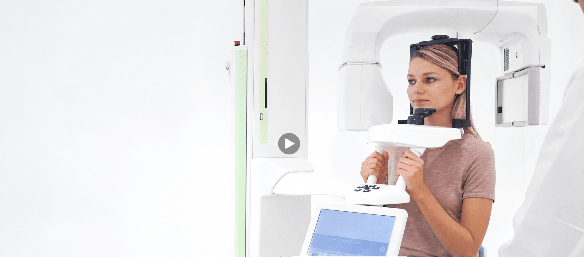
Planmeca 3D CT: understanding your anatomy
3D CT, also known as CBCT, shows the bone, roots, maxillary sinuses and the course of the nerve canals. These images help us assess the available space and identify structures that need protection during surgery.
The examination uses X-rays. Planmeca low-dose protocols tailor the scan to the area and the information required for diagnosis.
For you, this means a three-dimensional anatomical assessment carried out at the clinic.
3Shape TRIOS scanner: a digital impression
The intraoral scanner records your teeth, gums and bite and converts them into a three-dimensional model of the dental arches. During scanning, software combines hundreds of images to reproduce the condition of your mouth.
Scanning avoids traditional impression materials and lets us check the quality of the recorded information immediately on screen.
For you, this means a more comfortable way to take impressions and a digital model available to the team throughout treatment.
Digital Smile Design:
preview before deciding
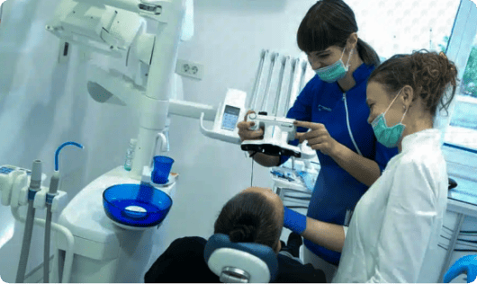
Photographs, intraoral scans and artificial intelligence technologies are combined to create a simulation of the planned smile after treatment.
We assess tooth shape, proportions and position in relation to your face, lips and expression. An in-mouth mock-up then allows you to assess the design and discuss any changes before treatment begins.
You can understand the proposed result and take part in the design decisions before treatment begins.
-
Dental lasers:
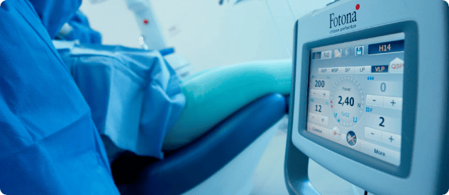
targeted treatmentA dental laser is selected according to the tissue and procedure. It may be used on teeth, gums and bone to remove decay, contour tissues or support root canal disinfection. It also has a role in specific surgical, periodontal, peri-implantitis and whitening protocols. When treating infections, it complements other necessary procedures rather than replacing them.
The clinician selects the laser and its application according to the tissue and procedure being treated.
-
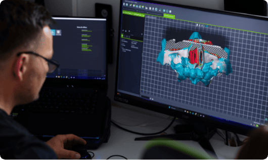
Computer-guided implant dentistry:
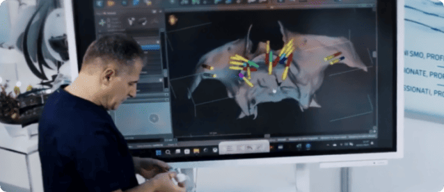
planning before surgery3D CT and intraoral scans are combined to define implant position, angle and depth in advance. Planning also considers the fixed teeth the implants will support.
Where indicated, a surgical guide made from the digital plan helps reproduce the planned implant position during surgery.
Surgery is planned around your anatomy and, where clinical conditions allow, may use a less invasive approach.
All technologies within one clinic
Diagnosis, scanning, planning and treatment are coordinated within the same clinic. The professionals involved can compare information directly and explain how each stage contributes to your care.
The examinations described here can be carried out at the clinic. Once treatment is planned, you will know the expected appointments and approximate duration of each stage, helping you organise your time.

Frequently asked questions
Does a 3D CT scan use radiation?
Yes. 3D CT, or CBCT, uses X-rays to produce three-dimensional images of bone, roots, nerve canals and maxillary sinuses. Exposure depends on the area being examined and the information needed for diagnosis. Planmeca low-dose protocols tailor the scan to the particular examination. A 3D CT scan is therefore not performed automatically: it is prescribed when three-dimensional information is useful for planning a surgical, implant or diagnostic procedure.
Are 3D CT and an intraoral scanner the same thing?
No. They collect different information. 3D CT uses X-rays to show internal structures such as bone, roots, nerve canals and sinuses. An intraoral scanner records tooth and gum surfaces and the relationship between the arches to create a three-dimensional digital model. Where needed, the two datasets are combined to plan treatment around your actual anatomy.
Does computer-guided implant surgery always avoid incisions and stitches?
No. Computer planning uses 3D CT and intraoral scans to define implant position, angle and depth in advance. In suitable cases, a surgical guide helps reproduce the planned position and may allow less invasive access. Incisions and stitches cannot always be avoided; this depends on anatomy, gums, available bone and the procedures actually required.
Does a dental laser always replace the drill or scalpel?
No. A laser is selected according to the tissue and treatment objective. It may be used on teeth, gums or bone, for example to remove decay, contour tissues or support canal disinfection. It is also used in certain surgical and periodontal protocols. It does not automatically replace conventional instruments; your clinician chooses tools according to the clinical situation.
Does Digital Smile Design show the exact final result?
Digital Smile Design presents a proposed smile for discussion before treatment. The simulation does not guarantee an identical final result. Photographs, scans and facial information help assess tooth shape, proportions and position. A temporary try-in can help you evaluate the proposal in your mouth. The final result depends on clinical conditions and the treatment performed.
Complete the form to book your free consultation with the Smile team.
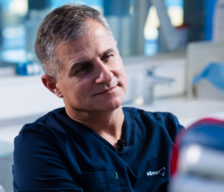
Darko Slovša and the Smile team
Specialist expertise, coordinated care.
Dr Darko Slovša, Dr. Sc., M.Sc., DDS, is a specialist in implantology and oral surgery. The Smile team brings together surgeons, dentists, dental technicians and assistants who work together throughout your treatment.